Watch the malaria parasite invade a human red blood cell. It takes 30 seconds.
(via New Scientist blog)
Middle and High School … from a Montessori Point of View
Watch the malaria parasite invade a human red blood cell. It takes 30 seconds.
(via New Scientist blog)


Well, since certain organelles within our cells (mitochondria) have their own DNA, it’s been suggested that they were once separate organisms that became the ultimate symbionts. Now, someone’s found that single celled amoebas may actually farm the bacteria they eat.
P.S. While looking for a picture of the guilty party, I came across this nice image of the amoeba, Dictyostelium discoideum, splitting into two on Wikimedia Commons.

The temperature dropped below zero (Celcius) for the first time last Friday night. We’d put back up the greenhouse’s plastic cover, which had blown off a couple weeks ago in a wind storm, but that was not enough to save a couple tomatoes and a squash plant.
It was a good illustration of the effects of freezing on plants not adapted to the colder weather. The leaves all turned black and flopped over, probably because the expanding ice ruptured the cell walls.
It also indicates that I need to get at temperature data-logger so I can monitor the temperature inside and outside the greenhouse. In the spring I hope to start a bunch of plants inside but put them into the greenhouse at the first opportunity, but I’ll need to make sure that greenhouse can support them. The data logger will also allow for some interesting experiments.
I’m just testing out a simple image map created with the GIMP. The GIMP is a free image manipulation software, a bit like Photoshop, not quite as sophisticated, but free. I used GimpTalk‘s very helpful guide. I though it would be easiest if I used something from a previous post as a test.
You should be able to click on the cell walls, chloroplasts, vacuole and nucleus. The links take you to the associated Wikipedia pages, but that’s just because this is a quick and dirty example. Image maps have been around for a long time, but I believe this is the first time I’ve ever created one. Now I just need to animate it a bit.
Unfortunately, this image is not easily scalable, though it should not be too hard to find (or write) a script to do just that.

One way to represent the process of mitosis is through dance. One of my students suggested they do an interpretive dance for their natural world personal project. I think they were mostly kidding, but with a fair bit of encouragement they did end up doing it.
The dance is much more literal than it probably needs to be since I helped a bit with the final product. I still think it’s pretty useful though because it’s abstract enough that you have to know the mitosis process to figure out what’s going on. So much so, I had them perform it twice at the end of our synthesis discussion. The second time through I narrated it so the steps would be clear to everyone.
I think it might make for a good “spark the imagination” lesson if one was needed.
Right now the dance needs four people, two for the chromosomes and two for the centrioles, but it would be really neat if the entire class participated by representing the cell membrane.
The diagram with the steps is: mitosis.svg. The instructions are below.


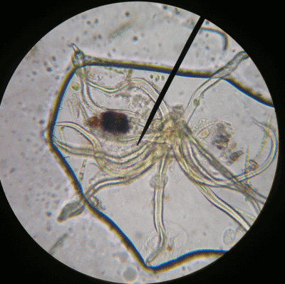
We spent the afternoon period on science. I’d given some individual microscopy lessons during the last immersion, where we looked at exciting protozoans moving around in pond water. This time they tried their hands at onion cells and staining with iodine, using a very nice and clear YouTube video posted below (kyliefansunited, 2008) as a reference.

The immersion oil had arrived in the mail earlier in the week so we got to try out the 100x oil lenses. We can now see structures inside the nucleus quite nicely.
Other things did not go so well. I’d written up, using the excellent recommendation of Anna Clarke, what I though was a neat exercise to look at the effect of osmosis on the cells of a waterplant, Egeria densa. The small group struggled with it, I think in large part because they were not quite prepared (had not done the background reading), and weren’t working very well together today. I’ll keep it on the schedule, but next time I’ll have to think hard on if it will be necessary to tweak the exercise.

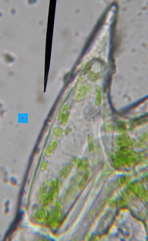
In a bit of a hurry, I swung by the pet store and picked up the aquatic water plant with the thinnest leaves I could find. It turned out to be Egeria densa, and while not the Elodea recommended by my expert contact Anna Clarke as a good subject for some microscope work, it seemed quite similar.

I needed the plant for an osmosis experiment. Dropping a little salt water on leaf cells of a freshwater plant should suck all the water out of the vacuoles and through the cell walls, potentially collapsing the cells (wouldn’t that be cool). I’d never done this before so I was quite curious to see what would actually happen.

The leaves have multiple layers of cells, so it’s hard to distinguish much at the center of a freshly clipped leaf, especially at high magnification. But if you look at the cells at the edges of the leaves, you can see some really neat looking, spiky cells, for which, I’m willing to bet, biologists have some really cool, multisyllabic name.

With a little bit of immersion oil and a 1000x objective, the spiky cells are good subjects for magnification: they’re a bit larger than their neighbors so they’re easier to see; their chloroplasts are distinct; and you can even make out the nucleus without staining.
Then I added the salt solution, and while the cell walls stayed strong, the cytoplasm collapsed into a little droplet at the center of the cell. The chloroplasts and the nucleus were all bundled together in this central blob (see the image at the top of the post). It’s quite the neat effect, though not exactly what I thought to see.

An interesting side note is that the cell nuclei show up very nicely with iodine stain, but the stain also discolors the chloroplasts.

One of our small group activities is to look at mitosis in onion cells. Anna Clarke, recommended the University of Arizona site which has an Online Onion Root Tips activity for those without access to the slides or microscope. It also provides a good review even if you do have those resources. Dr. Paul’s page on onion cell division is a good supplement to the Arizona site because of its great cell images.
If you’re feeling ambitious and want students to make their own slides, you can try the SAPS page on Mitosis in root tips.