She’s got (a) heart: Duck Dissection

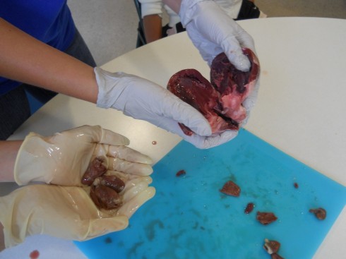
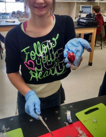
Heart in hand: a duck's heart to be precise.
Heart in hand: a duck’s heart to be precise.

Dissection time is always interesting in the middle school classroom. Some kids start off a little squeamish, but there are always a few who pitch right in. All of the class were working on different organisms–microscopic algae (desmids), insects, plants, etc.–but I set up the dissection table in the middle of the room. With all the other tables facing inwards, everyone who wanted to got a chance to see the internal organs as they were carefully, but triumphantly, traced and extracted. It spreads the enthusiasm, and gives the other students a heads up as to what will be expected.

One student brought in a duck that her father had shot. She was familiar with cleaning hunted fowl, so had no qualms about the dissection. The duck was a great specimen, because the internal organs were large and easy to identify.
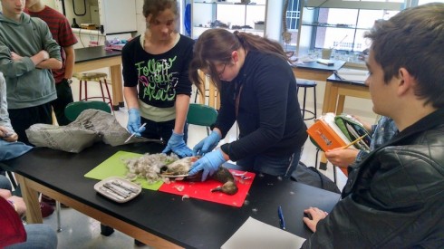
Duck dissection.
C.F. leads the duck dissection.

She, and most of the rest of the class, had dissected a sheep’s heart in elementary school. This time, however, they got to see how the heart was connected to the rest of the body. We got to talk about why smaller organisms, like ducks, could get away with two chambered hearts, while larger ones need four chambers.

Indeed, the tracing of the artery leading out of the heart lead us to compare of the color of the duck’s lungs–so red they were almost black–and the fish’s gills, which were also a deep red. And that, in turn lead to a discussion of how organisms ingest the gasses they need to survive: microbes through diffusion; insects through spiracles and trachea; mammals through lungs. Tomorrow we’ll talk about how the surface area to volume ratio changes with size.
Rabbit Dissection

Yesterday, Dr. Sansone was kind enough to lead my Biology class through a small mammal dissection (details here). He’d brought in five New Zealand White rabbits (2 male and 3 female) that had been raised for market by an Amish family in northern Missouri.

Over our two-hour class period, we had time to examine the organs in the abdominal and thoracic cavities in some detail. Students had been prepared with group reading assignments of the different organ systems based on the anatomy outline.

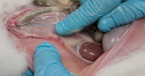
Dr. Sansone points out a rabbit's ovaries. A kidney is visible to the lower right.
Dr. Sansone points out a rabbit’s ovaries. A kidney is visible to the lower right.

After the dissection we removed the pelts and froze them for later preservation. We also froze the carcases for later. I’ve convinced Mr. E. that cooking them would be a great interim activity. Mr. E. is a bit of an epicure, so it did not take much convincing.

Notes

Only one student declined to participate in the dissection for ethical reasons. About half of the class declined to observe the harvesting. Four students volunteered to assist in the harvesting, two of whom were not even in my biology class. They were seniors. One of them, P., had done it before and demonstrated the procedure for us.

An Outline for Anatomy

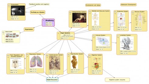
Concept map for the study of anatomy.
Concept map for the study of anatomy. This diagram will be used for the introductory lesson.

Because we have the opportunity to get a few rabbits, we’re starting Biology this year with anatomy. In the first week, our discussions will be based around the rabbit dissection so we’ll be focusing on the systems where the organs are easiest to find: the fluid regulation/excretory, respiratory, cardiovascular, reproductive, and digestive systems.

Preparing for Rabbit Dissection

References

Linn (2000) has a good overview of general rabbit biology and the internal organs in Rabbits: Biology (pdf).

For lots of detail on how to dissect a rabbit try Bensley’s Practical Anatomy of the Rabbit (free on Google Books)

AnimalLearn.org has a good list of free and for fee online dissections of various organisms. No rabbits as of this moment though.

Drawing Daffodils

It’s spring, and what better time to study meiosis and dissect daffodils.

Students collect daffodils for dissection.

Daffodilusa (pdf) has nice description of how to dissect daffodils. However, I had students collect the flowers, and sketch the outsides and insides (longitudinal bisection) before I gave them the handout.

I wanted them to practice drawing diagrams and observing features first, before we got into the discussion of what the parts were and what they did, to make sure they’d not forgotten all they’d learned when we did our animal dissections last semester.

They laid out their grids, did some very nice drawings, and then labeled what they’d drawn, based on the handout, over the weekend.

Shrimps are not for Wimps

Shrimps are not for Wimps
by Abby Reynolds

One day I walked into the science class,
I was preoccupied as I looked into the looking glass.
For Dr. Urbano had cooked up a surprise,
And after I wished I was at the car rental Enterprise.
Then I would be able to drive away,
From the disgusting horrors I saw that day.
It started with a metal tray,
With icky rubber a sickly gray.
He brought the probe and scalpel too,
My o my, Sydney turned blue!
Then a smelly smell filled the room,
Making us cough and gag all too soon.
When I saw the shrimp my stomach did flip-flop,
For the color was like that of a caramel lollipop.
I inhaled through my mouth to calm my brain,
Trying not to think how the shrimp was slain.
It helped somewhat until I saw Sydney,
Who was slicing and dicing at a kidney.
She looked up and grinned a big grin,
A smear of blood dripping down her chin.
I will kindly spare you the rest of the story,
For I fear that it gets MUCH too gory!

And there I was thinking that the shrimp dissection had gone rather well.

Shrimp

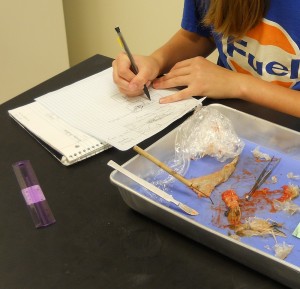
Drawing an external diagram of a jumbo shrimp.

Our middle-school dissections have moved on from hearts to whole organisms. This week: jumbo shrimp.

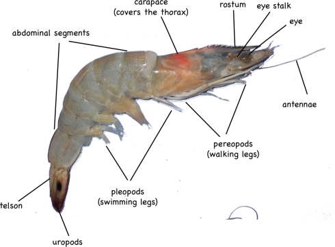
I particularly like these decapods because the external anatomy is simple but interesting, including: eyes on stalks; a segmented body; 5 pairs of swimming legs; 5 pairs of walking legs. The simple, clear layout make them a good subject for students to work on improving the accuracy of their full-scale drawings.

External anatomy of a shrimp.

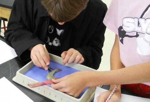
The internal anatomy is a bit harder to distinguish, however, since the organs are relatively small. Most of my students found it difficult to remove the carapace without smushing everything inside the thorax, which includes the stomach, heart, and digestive gland.

Dissecting the shrimp.

The abdominal segments were easy to slice through, on the other hand, and we were able to identify the hindgut (intestine), which runs the length of the back side, and the blueish-colored, nerve cord that is nearer the front (ventral) side.

Under the microscope, you could see little mineral grains in the contents of the gut. Although I did not manage to, I wanted to also mount the thin membrane beneath the carapace on a slide. If I had, we might have been able to see the chromatophores, “star- or amoeba-shaped pigment-containing organs capable of changing the color of the integument” (Fox, 2001).

References

Richard Fox (2001) has a good reference diagram and description of brown shrimp anatomy.

M. Tavares, has compiled some very detailed shrimp diagrams (pdf) (originally from Ptrez Farfante and Kensley, 1997)

Hearts

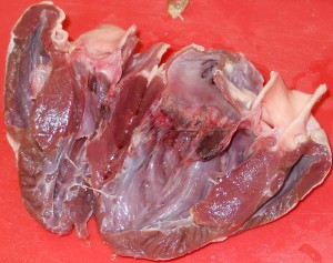
Chicken hearts (left) and a pig heart (right).

My middle school class has been looking at organ systems and we’ve started doing a few dissections. We compared chicken and pig hearts last week.

Pig heart.

Pig hearts are large and four-chambered like ours, so they should have matched up very well with the diagrams from the textbook. However, real life tends to be messy, which is one of the first lessons of dissection. It was tricky finding all the chambers, and identifying the valves, even when you knew what you’re supposed to be looking for. It is especially difficult, as one of my students noted, because everything isn’t color coded.

Chicken hearts are a bit trickier, because they’re a lot smaller. They also have four chambers, but the main chamber (the left ventricle) is so dominant that it’s easy to assume that there’s only the two (or even just one) chambers.

One student’s interesting observation was that the pig heart was a lot more pliable than the chicken heart. My best guess was that the chicken heart is made of a tougher muscle — it’s denser and more elastic (in that it rebounds to its original shape faster) — because has more work to do: chicken heart rates can get up to 400 beats per minute (Swinn-Hanlon, 1998) compared to 70 beats per minute for the pig.

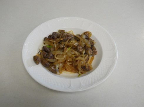
To get a better feel for the texture, and to engage our other senses in our observations on the hearts, at the end of the dissection, which was conducted in the dining room using kitchen utensils, I fried up some of the chicken hearts with onions for lunch.

Notes

There are a number of nice labs for heart dissections online:

The hearts were purchased at the Chinese supermarket, Seafood City. There seems to be a greater organ selection on the weekends.