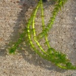
In a bit of a hurry, I swung by the pet store and picked up the aquatic water plant with the thinnest leaves I could find. It turned out to be Egeria densa, and while not the Elodea recommended by my expert contact Anna Clarke as a good subject for some microscope work, it seemed quite similar.
I needed the plant for an osmosis experiment. Dropping a little salt water on leaf cells of a freshwater plant should suck all the water out of the vacuoles and through the cell walls, potentially collapsing the cells (wouldn’t that be cool). I’d never done this before so I was quite curious to see what would actually happen.
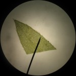
The leaves have multiple layers of cells, so it’s hard to distinguish much at the center of a freshly clipped leaf, especially at high magnification. But if you look at the cells at the edges of the leaves, you can see some really neat looking, spiky cells, for which, I’m willing to bet, biologists have some really cool, multisyllabic name.
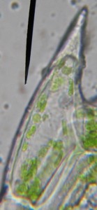
With a little bit of immersion oil and a 1000x objective, the spiky cells are good subjects for magnification: they’re a bit larger than their neighbors so they’re easier to see; their chloroplasts are distinct; and you can even make out the nucleus without staining.
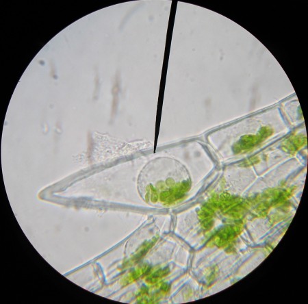
Then I added the salt solution, and while the cell walls stayed strong, the cytoplasm collapsed into a little droplet at the center of the cell. The chloroplasts and the nucleus were all bundled together in this central blob (see the image at the top of the post). It’s quite the neat effect, though not exactly what I thought to see.
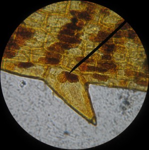
An interesting side note is that the cell nuclei show up very nicely with iodine stain, but the stain also discolors the chloroplasts.