Cells, cells, cells

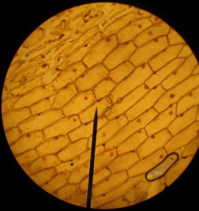
Onion cells stained with iodine. 100x magnification.

We spent the afternoon period on science. I’d given some individual microscopy lessons during the last immersion, where we looked at exciting protozoans moving around in pond water. This time they tried their hands at onion cells and staining with iodine, using a very nice and clear YouTube video posted below (kyliefansunited, 2008) as a reference.

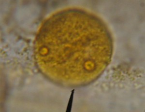
Nucleus of an onion cell stained with iodine and, for experimentation, Congo Red. 1000x magnification

The immersion oil had arrived in the mail earlier in the week so we got to try out the 100x oil lenses. We can now see structures inside the nucleus quite nicely.

Other things did not go so well. I’d written up, using the excellent recommendation of Anna Clarke, what I though was a neat exercise to look at the effect of osmosis on the cells of a waterplant, Egeria densa. The small group struggled with it, I think in large part because they were not quite prepared (had not done the background reading), and weren’t working very well together today. I’ll keep it on the schedule, but next time I’ll have to think hard on if it will be necessary to tweak the exercise.

Osmosis under the microscope

The effects of placing freshwater plant cells (Egeria densa) in salt water solution.

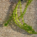
In a bit of a hurry, I swung by the pet store and picked up the aquatic water plant with the thinnest leaves I could find. It turned out to be Egeria densa, and while not the Elodea recommended by my expert contact Anna Clarke as a good subject for some microscope work, it seemed quite similar.

Egeria densa plants sitting in shallow water in the sun.

I needed the plant for an osmosis experiment. Dropping a little salt water on leaf cells of a freshwater plant should suck all the water out of the vacuoles and through the cell walls, potentially collapsing the cells (wouldn’t that be cool). I’d never done this before so I was quite curious to see what would actually happen.

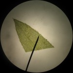
Leaf tip of Egeria densa. 40x magnification.

The leaves have multiple layers of cells, so it’s hard to distinguish much at the center of a freshly clipped leaf, especially at high magnification. But if you look at the cells at the edges of the leaves, you can see some really neat looking, spiky cells, for which, I’m willing to bet, biologists have some really cool, multisyllabic name.

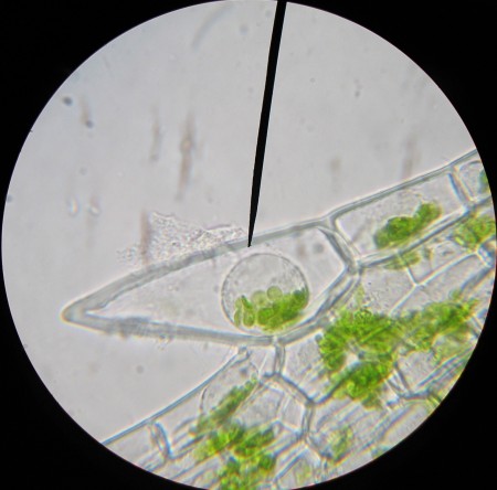
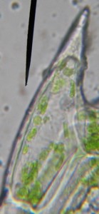
Spiky cell under 1000x magnification.

With a little bit of immersion oil and a 1000x objective, the spiky cells are good subjects for magnification: they’re a bit larger than their neighbors so they’re easier to see; their chloroplasts are distinct; and you can even make out the nucleus without staining.

Then I added the salt solution, and while the cell walls stayed strong, the cytoplasm collapsed into a little droplet at the center of the cell. The chloroplasts and the nucleus were all bundled together in this central blob (see the image at the top of the post). It’s quite the neat effect, though not exactly what I thought to see.

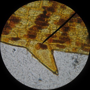
Spiky Egeria cells with iodine stain.

An interesting side note is that the cell nuclei show up very nicely with iodine stain, but the stain also discolors the chloroplasts.

Oven calibration

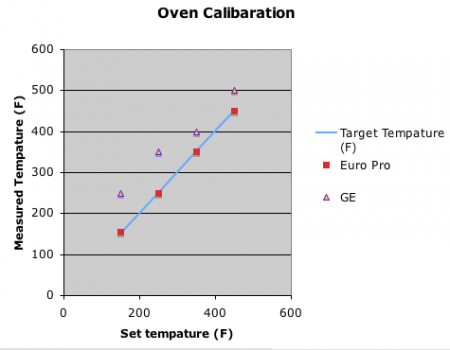
Initial oven calibration curves (2009).

Catastrophic failure of one of our ovens! Last year when we started up the bread business, we bought two counter-top ovens within a couple of weeks of each other. They needed to be extra-large to fit two loaves of bread each, which made them a little hard to find. We got a EuroPro oven first, and when we found that it worked pretty well, we went back to try to get another. But just a week later, the store was out of stock and that type of oven could not be found in the city of Memphis or its environs.

Instead we got a GE model. The price was about the same, as was the capacity. We quickly realized that the GE was quite the inferior product. The temperature in the oven was never the same as what was set on the dial. Our bread supervisor at the time ran a calibration experiment, the results of which you can see above, so we still managed to use the oven. Only this year, three weeks into the term, it conked out.

We sold at least one underdone loaf before we realized what had happened, and received a detailed letter in response (which our current bread supervisor handled wonderfully in his own well worded letter). Fortunately, we have found a newer version of our EuroPro oven, which seems to work quite well.

I like the oven calibration exercise. It was a nice application of the scientific process to solve an actual problem we had with the business. Though I know it’s not quite the same, I like the idea of doing annual oven calibrations just to check the health of our equipment and help students realize that the scientific process is a powerful way of looking at the world, not just something you do in science.

More mitosis resources

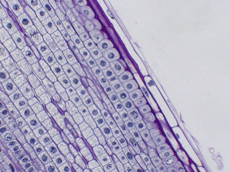
Onion root tip cells, with chromosomes stained to show cells in different stages of mitosis. Image from uafcde.

One of our small group activities is to look at mitosis in onion cells. Anna Clarke, recommended the University of Arizona site which has an Online Onion Root Tips activity for those without access to the slides or microscope. It also provides a good review even if you do have those resources. Dr. Paul’s page on onion cell division is a good supplement to the Arizona site because of its great cell images.

If you’re feeling ambitious and want students to make their own slides, you can try the SAPS page on Mitosis in root tips.

Mitosis resources

Our assignments for natural world usually combine some reading and some type of activity, but all the short video clips available online are a great resource, so I’ve been adding them to the studyguides as I find them.

The above two-minute, cell division video is a great example. Mitosis is a process, so it makes a lot of sense showing it as an animation, rather than discrete pictures in a figure. The video makes deciphering what’s going on in the diagram in the textbook a whole lot easier to understand, while the textbook diagram fills in the detail so the whole thing makes more sense.

There are also a number of useful interactive animations online. John Kyrk’s is quite nice. I like how the CellsAlive animal cell mitosis page lets you step through each frame in the animation.

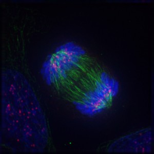
Anaphase: Lengthening microtubules push the two sets of chromosomes further apart. (from Wikipedia)

Wikipedia, as is so often the case, also has some nice images.

Spore: Lamarkian in the subtexts

Playing Spore.

During our last immersion, one of my students brought in the computer game, Spore. Although the game subtly indicates that it’s your progeny that gains evolutionary advances, the fact that you get to choose what you want (extra horns, poison sacks), and the fact that you can see yourself (or do I mean your creature) evolve on the screen, really smacks of Lamarckism. While it’s appealing to think, like Lamark, that you can pass on traits gained during your lifetime to your kids, despite some fascinating new research, that’s just not how evolution works.

Evolution is not directed by the organism but by their environment. In a population of organisms of any particular species there is going to be some variability due to simple, random genetic mutation. Some lucky members of the species might have a mutation that makes their muscles better at burning oxygen during sprints, making them able to run faster to get away from the lions. So they survive and pass their genes on, with their genetic mutation. Of course, if lions become extinct (disease maybe) then this trait may no longer be beneficial and something else, like maybe intelligence, would be selected for.

The game can capture your interest, however, so I’ve asked the student who brought in the game to come up with a presentation explaining why it would be useful to have the game in the classroom. I am, after all, not instinctively opposed to using computer games in class. I’m really curious to see what this game looks like from the student’s point of view.

Jurassic Park: Web of Issues

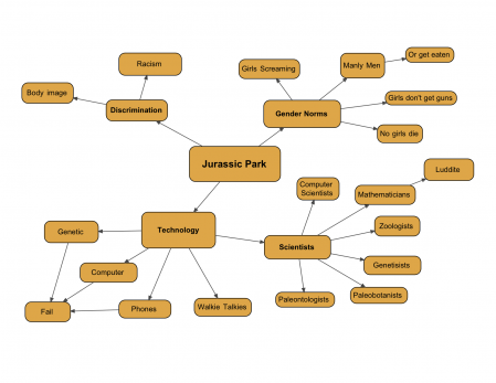
Web of issues for the movie Jurassic Park.

Well we watched Jurassic Park last night and concluded it with a discussion about the issues underlying the movie, the same way we’ve been studying analyzing the issues underlying texts. Discrimination based on race and obesity came up first (the fat guy and the black people “always” die), but I was able to coax a bit of discussion about the role and responsibility of science and scientists. Our discussion is summarized in the graphic organizer above, but there are many more subtexts to the story that we did not have time to explore.

Jurassic Park by Michael Crichton (the book).

I like both the movie and the book because, like most good science fiction, they explore some interesting issues that relate quite nicely to the curriculum. Jurassic Park has a nice little introduction to DNA and gene sequencing that is tied to some the history of life on Earth. As works of art in their respective fields, however, I prefer the movie. The novel has a lot of wonderful detail, and the scientist in me loves the detail, but the characters are not as well drawn and the story seldom strays from its main thesis, scientific hubris. What it has to say about that issue is well expressed and well researched so it does capture the interest of the reader. (The follow-up book, “The Lost World”, sails adrift of the science, is logically incoherent and has a proportionate deterioration in the quality of the writing.) I do however recommend the original Jurassic Park book to my students as a personal novel.

Steven Spielberg makes a great movie, extracting empathetic performances from the actors. Since the book’s author, Michael Crichton, also wrote the screenplay, the movie stays true to the core issues in the text. I think its a great example of a successful, dare I say synergistic, collaboration.

Tomorrow, instead of retelling around the issues in writing, my students are going to try to do so in a skit. This could get interesting.

Microscope photography!!!

Algae and amoebas at 400 times magnification.

Did you know that if you hold up a regular digital camera up to the eyepiece of a microscope you can take a great picture of a magnified slide! I didn’t. And I really didn’t think it would work when I tried it, but the results are remarkable. With a somewhat steady hand you can also make decent animations.

If you look carefully you can see the amoebas zipping around. I also have a really cool larger version too, which shows the entire slide..

I’ve never been very good at identifying things (I’m a lumper not a splitter) so all I think I can say for sure is that there are algae and protozoans in the picture. BiologyCorner has a nice identification guide for organisms usually found in ponds, which is part of one of their lessons, Biodiversity of Ponds.