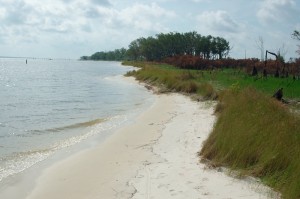
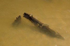
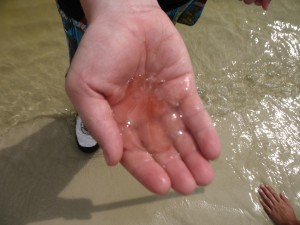
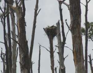
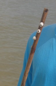
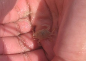
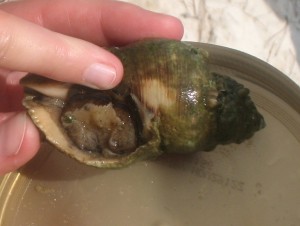
We were there to collect garbage, but we found lots of life on the Deer Island part of our adventure trip to the gulf.

Middle and High School … from a Montessori Point of View

We were there to collect garbage, but we found lots of life on the Deer Island part of our adventure trip to the gulf.


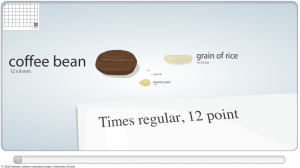
The Genetic Science Learning Center (which I’ve mentioned before) has a wonderful slider-bar animation that shows the differences in scale from what we perceive (a grain of rice or a coffee bean) down to the scale of cells, molecules and finally a carbon atom.
The smallest objects that the unaided human eye can see are about 0.1 mm long. That means that under the right conditions, you might be able to see an ameoba proteus, a human egg, and a paramecium without using magnification. …
Smaller cells are easily visible under a light microscope. It’s even possible to make out structures within the cell, such as the nucleus, mitochondria and chloroplasts. [my example] … The most powerful light microscopes can resolve bacteria but not viruses.
To see anything smaller than 500 nm, you will need an electron microscope. … The most powerful electron microscopes can resolve molecules and even individual atoms.
–Genetic Science Learning Center (2011, January 24) Cell Size and Scale. Learn.Genetics. Retrieved June 13, 2011, from http://learn.genetics.utah.edu/content/begin/cells/scale/
The … organic materials in [some] meteorites probably originally formed in the interstellar medium and/or the solar protoplanetary disk, but was subsequently modified in the meteorites’ asteroidal parent bodies. … At least some molecules of prebiotic importance formed during the alteration.
Herd et al., 2011: Origin and Evolution of Prebiotic Organic Matter As Inferred from the Tagish Lake Meteorite
Amino acids are the building blocks of life as we know it. They can be formed from abiotic (non-biological) chemical reactions (in a jar with electricity for example). It’s been known for a while that amino acids can be found on comets and asteroids, but now this fascinating article suggests that a lot of the chemical reactions that created these precursors to life happened on the asteroids themselves. Then when the asteroids bombarded the Earth, the seeds of life were delivered. More details here.
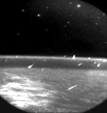
This, of course, is just one of several hypotheses about the origin of life on Earth: livescience.com outlines seven.
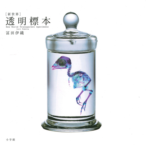
After the joy of playing around with microscopy and staining last fall, it’s no surprise that someone has taken the science of staining and specimen preservation and turned it into art.
Iori Tomita has done an amazing job at making visible the internal organs of the specimens.
Using a method that dissolves an animals natural proteins, Tomita is able to preserve these deceased animals with striking detail–highlighting the finest and most delicate skeleton structures.
To further enhance the visual appeal of these ornate skeletons, Tomita selectively injects different colored dies into hard bones and soft bones to create a 3-d effect. Without the addition of the dye, the animals remain translucent.
— Michael (2010): New World Transparent Specimens Turns Preservation Into Modern Art
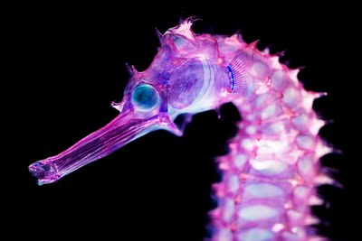
Tomita’s website has some excellent photographs, and there appear to be two books available from Amazon.com.jp. More pictures can be found online here and here. Lisa Stinson at Wired has more pictures and details on the method.

To follow up my own attempts at a fish anatomy lesson, I asked the people at the Gulf Coast Research Lab’s Marine Education Center to include a dissection in their program for our Adventure Trip. They chose squid.
Squid are nice because they’re mostly soft tissue and the organs are fairly easy to identify. They’re also quite charismatic, which piqued the students’ interest. These squid were going to be used as bait, so I didn’t feel too badly about using them for science.
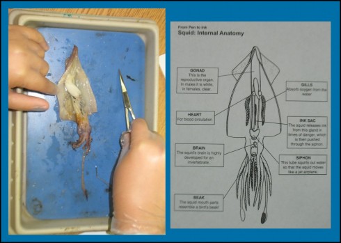
Once again, our guide, Stephanie, was an excellent teacher. A good time was had by all, even though it was a bit gruesome.
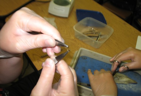
I would have liked to have a little more time to draw some diagrams, but I don’t think my students would have had the patience. It was the Adventure Trip after all, and they’d much rather spend the time outside.
As for the future, I like this note about squid dissection:
… this … is a tactile experience. You may want to explore this aspect through sensory activities, written descriptions, poetry, and/or artwork. Encourage students to experience the many textures found inside and outside the squid’s body. Moving fingertips along the suckers is suggested as well – the suckers do not scrape or hurt if you are gentle with them.
–Center for Educational and Training Technology, Mississippi State University: Squid Dissection
This quote comes from a Mississippi State website, which also has a great set of calamari recipes in addition to dissection instructions. I’m always in favor of an interdisciplinary approach; food-preparation rather than purely dissection.
Finally, the University of Buffalo’s Biology 200 class has some excellent, labeled pictures, for reference.
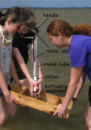
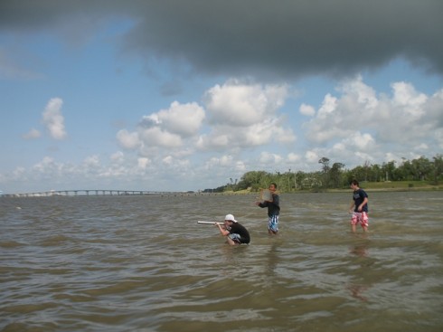
On the first morning of the Coastal Science Camp, between dip netting and seining at the estuary, we tried sampling beneath the seabed using a little coring device which I seem to have to forgotten the name of.
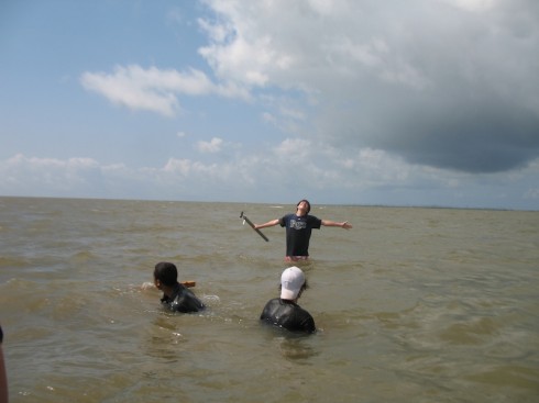
Usually, they can see the little holes in the seabed where the benthic macrofauna live, but not this time. All the sediment pouring into the Mississippi Sound from this spring’s swollen rivers had made the waters too turbid to see through. So we were coring blind.
The corer is simply a metal (stainless steel) barrel with a rubber piston inside. The piston is connected to a handle at the top with metal rod. To sample, you put the tip of the barrel at the sediment-water interface and push the barrel into the sediment at the same time holding the handle steady to keep the piston from moving into the sediment. Holding the piston steady provides a little suction on the inside of the barrel, which helps the barrel move into the sediment, and keeps the sediment in the barrel when you pull it out. However, it does help to put your hand on the bottom of the barrel as soon as possible to keep the sediment from falling out, even if that means sticking your hand into the sediment itself.
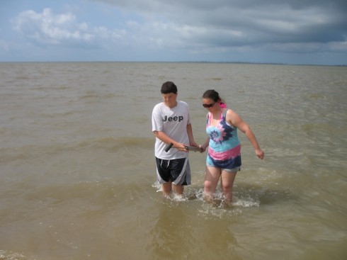

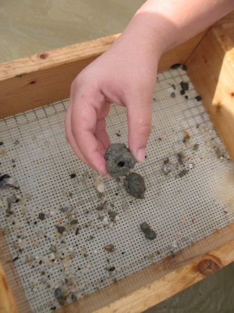
Once you’ve recovered the sediment, you extrude it into a sieve. Sometimes you can see a little layering in the extruding sediment, but we did not take the time to try to interpret it since our focus was on finding benthic fauna.
The sieve’s mesh is pretty coarse, so anything sand sized or smaller is washed out as you gently rock it back and forth in the water. We did not find much. Mostly small pebbles. Without being able to see the seabed our sampling pattern was pretty random.
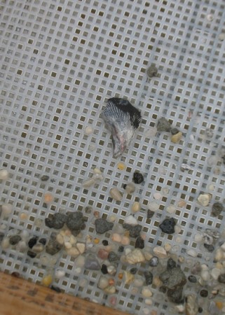
The more persistent groups (the class had been broken into groups of two or three) did find a couple things, including a polychaete, which is a segmented worm.
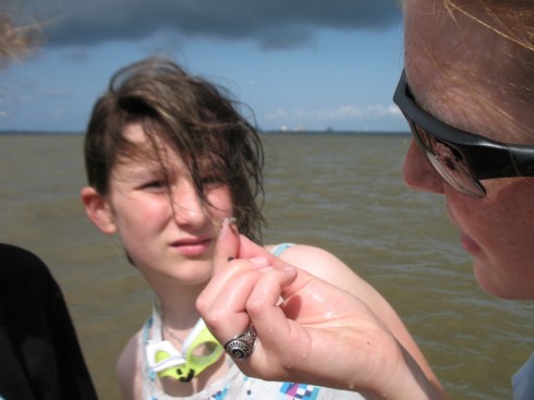
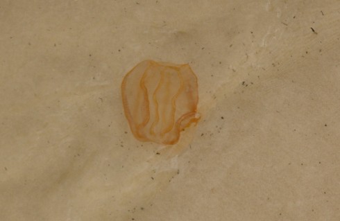
They also turned up a small, clawed, lobster-like organism:
We also found the burrow of an unknown organism, surrounded by a clayey cast. It looked very much like some of the fossilized burrow casts we saw at Coon Creek.
This type of sampling was not everyone’s cup of tea, however. Fortunately, the water was shallow and warm, so a good time was had by all.
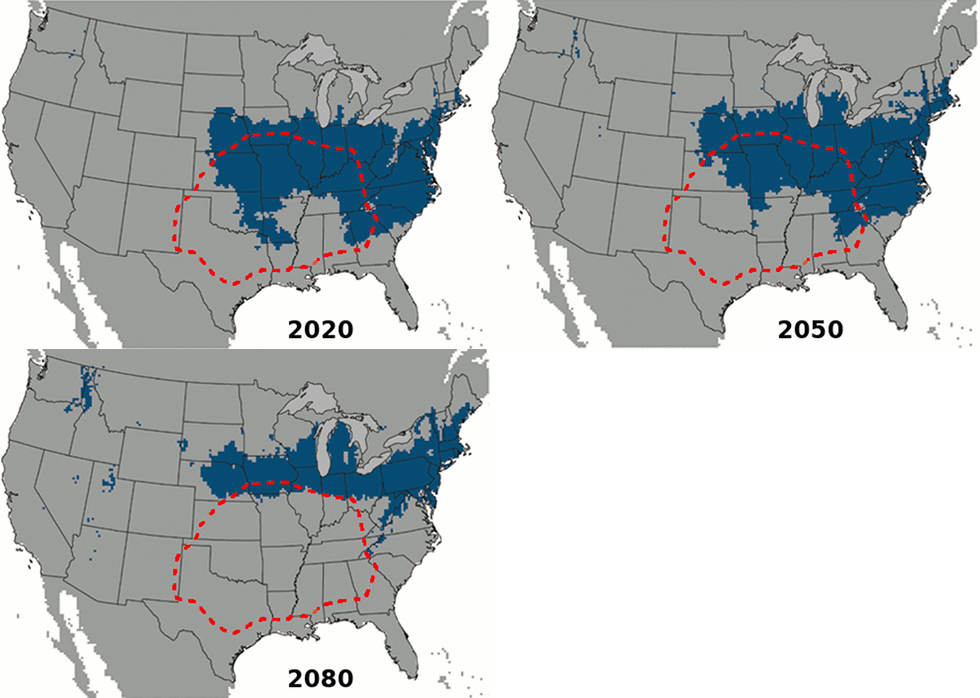
Just in time for us to learn about global change, this interesting study on the expanding range of brown recluse spiders came out. Once restricted to the southern U.S. and the midwest, future climate change will allow them to expand north to Minnesota and east into Pennsylvania.
The researchers, Saupe et al. (2011), used ecological niche modeling. This method takes known information about where the spiders live, such as climate (e.g. summer temperatures) or topography (e.g. mountains versus plains), to figure out the current extent of their ecological niche. Then they use climate models to figure out where those same conditions will apply in the future. Thus the spiders march north.
First off, three cheers for Viet Hoa, the Vietnamese food market on Cleveland Ave in Memphis. I’ve been searching for a source of fresh, uneviscerated, marine fish in this mid-continental city for quite a while, and I’ve finally found them. The market was the source of the subjects of an excellent anatomy lesson, and a delicious dinner.
I’d grabbed three fish, and a few goodies, and packed them on ice on the day before our trip. The large red snapper was a little pricey because it was fairly big, but I knew it would be quite tasty and I was hoping the internal organs would be big and clearly visible. The milk fish was an unknown, but it was big and cheap so worth experimenting on. The last fish was the smallest, and I’m not quite sure what kind it is, except that it’s marine. I’d tried one at home the previous week and found that while it had an excellent taste, the internal bits were on the small side.
On the first night of the immersion I started with the biggest fish, the red snapper, with everyone around the table. I’m happy to say that all my students were there for the lesson, facing the table, even the more vegetarian minded. I’ve always believed that there is a certain, necessary, ethic to knowing where your food comes from, and what went into preparing it for you. If you’re going to eat meat, you should be able to spare a moment to think about the animal.

The digestive system was the easiest to identify and trace. The mouth and anus are pretty obvious on the outside. After cutting through the skin of the belly, you can follow the intestines from the anus all the way to the stomach. The intestines are quite long.
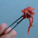
We found two partially digested, shrimp-like creatures in the stomach of the red snapper.
From the other end of the digestive system, once you get all the contents out of the body cavity, you can stick a probe through the mouth (watch out for sharp teeth) down through the esophagus to the stomach.
Pulling back the gills, you can see all the way through to the mouth. One student noticed how bright red the gills were, so we talked about oxygenation of blood and compared the fish’s gills to the human respiratory system.
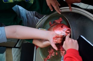
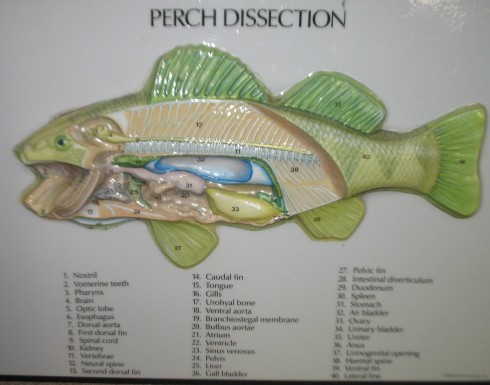
Judging from the fish anatomy diagram on the La Crosse Fish Health Center website, we found the air bladder (tucked in the center right next to the spine), the liver, the kidneys, and probably the heart. We pulled them out and photographed them.
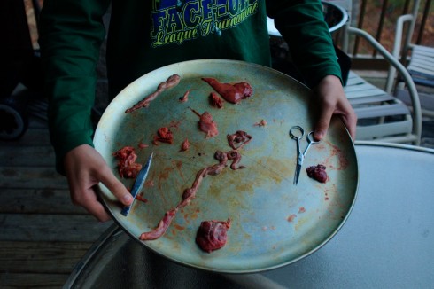
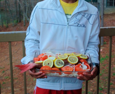
Finally, I laid the fish on a bed of thinly sliced onions, surrounded it with wedges of tomato (seeded), covered it with lemon slices, dribbled a tablespoon of olive oil on top, and forgot to add the cup of white wine (which we did not seem to have on hand for some reason) and the bay leaf. It was a big fish so we ended up baking it for about 40 minutes at 400 degrees Fahrenheit.
Then we ate it. I say we, but it really was just a couple of students and myself who did most of the eating, although I believe I convinced everyone to at least try it. Of the more serious students of anatomy, the eyeballs were highly prized; with only three fish we did not have enough to go around.
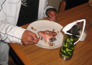
By the time we were done with dinner the skeletal system was very nicely exposed.
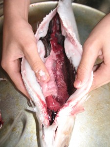
A couple of my students did the honors of cleaning the other two fish. With its large organs, the milk fish was an excellent subject for dissection, but it was not particularly palatable. We did however find the gall bladder.
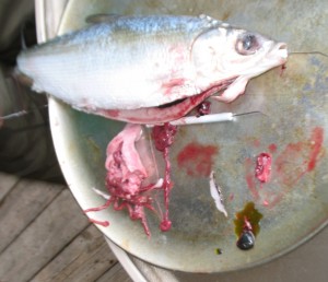
Even the smallest fish proved a worthwhile subject for the more patient student.
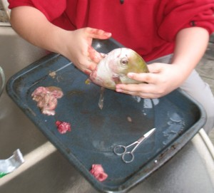
Instead of eating the rather unappetizing, baked milk fish I combined it in a soup with some clams I’d picked up from Viet Hoa. I’d grabbed the clams so we compare modern bivalve shells to the cretaceous ones we’d find at Coon Creek. A bit of boiling with a few herbs, made for an excellent broth the following night.
Cleaning fish for an anatomy lesson worked very well. As we excavated each internal organ, we could talk about what it did, why the fish needed it, and what was its analogue in humans. And, in the end, they made for a couple excellent meals.
P.S. – From the Gulf Coast Research Lab’s Marine Education Center, here’s a reference image for a perch dissection.
