Shrimp

Drawing an external diagram of a jumbo shrimp.

Our middle-school dissections have moved on from hearts to whole organisms. This week: jumbo shrimp.

I particularly like these decapods because the external anatomy is simple but interesting, including: eyes on stalks; a segmented body; 5 pairs of swimming legs; 5 pairs of walking legs. The simple, clear layout make them a good subject for students to work on improving the accuracy of their full-scale drawings.
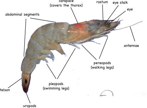
External anatomy of a shrimp.

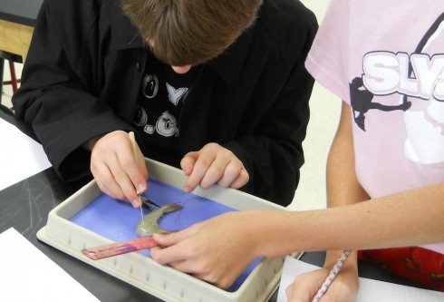
The internal anatomy is a bit harder to distinguish, however, since the organs are relatively small. Most of my students found it difficult to remove the carapace without smushing everything inside the thorax, which includes the stomach, heart, and digestive gland.
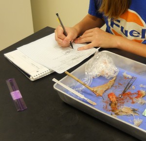
Dissecting the shrimp.

The abdominal segments were easy to slice through, on the other hand, and we were able to identify the hindgut (intestine), which runs the length of the back side, and the blueish-colored, nerve cord that is nearer the front (ventral) side.

Under the microscope, you could see little mineral grains in the contents of the gut. Although I did not manage to, I wanted to also mount the thin membrane beneath the carapace on a slide. If I had, we might have been able to see the chromatophores, “star- or amoeba-shaped pigment-containing organs capable of changing the color of the integument” (Fox, 2001).

References

Richard Fox (2001) has a good reference diagram and description of brown shrimp anatomy.

M. Tavares, has compiled some very detailed shrimp diagrams (pdf) (originally from Ptrez Farfante and Kensley, 1997)
Hearts

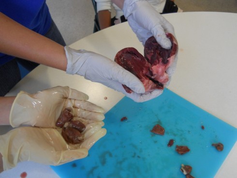
Chicken hearts (left) and a pig heart (right).

My middle school class has been looking at organ systems and we’ve started doing a few dissections. We compared chicken and pig hearts last week.

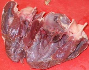
Pig heart.

Pig hearts are large and four-chambered like ours, so they should have matched up very well with the diagrams from the textbook. However, real life tends to be messy, which is one of the first lessons of dissection. It was tricky finding all the chambers, and identifying the valves, even when you knew what you’re supposed to be looking for. It is especially difficult, as one of my students noted, because everything isn’t color coded.

Chicken hearts are a bit trickier, because they’re a lot smaller. They also have four chambers, but the main chamber (the left ventricle) is so dominant that it’s easy to assume that there’s only the two (or even just one) chambers.

One student’s interesting observation was that the pig heart was a lot more pliable than the chicken heart. My best guess was that the chicken heart is made of a tougher muscle — it’s denser and more elastic (in that it rebounds to its original shape faster) — because has more work to do: chicken heart rates can get up to 400 beats per minute (Swinn-Hanlon, 1998) compared to 70 beats per minute for the pig.

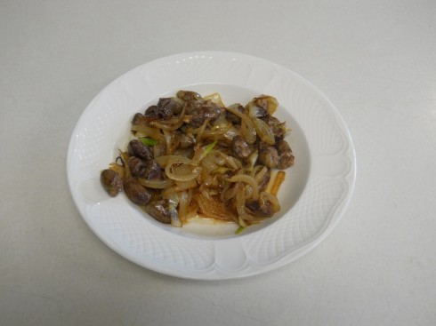
To get a better feel for the texture, and to engage our other senses in our observations on the hearts, at the end of the dissection, which was conducted in the dining room using kitchen utensils, I fried up some of the chicken hearts with onions for lunch.

Notes

There are a number of nice labs for heart dissections online:

The hearts were purchased at the Chinese supermarket, Seafood City. There seems to be a greater organ selection on the weekends.

Human Microbiome Project: Figuring out who our Microscopic Symbiotes are, and what they do

Although we are, by number at least, 90% bacteria, only recently have scientists with the Human Microbiome Project have just begun figuring out who those bugs are.

[The] trillions of microbes that make their homes in and on us do an excellent job keeping us healthy (crowding out harmful microbes) and sated (breaking down a lot of the food we ingest).

Now that disturbances in this rich microbiome community have been linked to weight gain, inflammatory bowel disease, vaginal infections and risk for infection with harmful microbes (such as methicillin-resistant Staphylococcus aureus, or MRSA), the importance of understanding what makes up a “healthy” microbiome has become even more apparent.

— Harmon (2012): Body Count: Taking Stock of All the Bugs That Call Humans Home in Scientific American.

On the Origin of Species

Perhaps the key reason for the profound influence of Darwin’s “On the Origin of Species” is that it’s such a well written and well reasoned argument based on years of study. It is a wonderful example of how science should be done, and how it should be presented. In the past I’ve had my middle schoolers try to translate sections of Darwin’s writing into plainer, more modern English, with some very good results. They pick up a lot of vocabulary, and are introduced to longer, more complex sentences that are, however, clearly written.

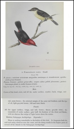
Diagram and notes on the bird species P.Nanus from The Zoology of the Voyage of H.M.S. Beagle, Part 3: Birds by J. Gould and G.R. Gray (edited by C.Darwin). Image via Darwin Online.

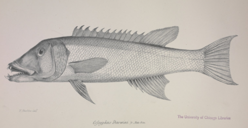
The text of “On the Origin of Species” is available for free from the Gutenberg library. Images of the original document can be found (also for free) at the UK website, Darwin Online (which also includes the Darwin’s annotated copy). Darwin Online also hosts lot of Darwin’s other works, as well as notes of the other scientists on The Beagle, among which is included some wonderful scientific diagrams.

This year, I’m going to have the middle schoolers read the introduction, while the honors environmental science students will read selected chapters and present to the class — this will be their off-block assignment.

Diagram of the fish Cofsyphus Darwini by L. Jenyns in The Zoology of the Voyage of H.M.S. Beagle, Part 4: Fish (edited by C.Darwin). Image via Darwin Online.. .

A Plant Responds to Water

Pepper plant responding to being watered. These images were taken over the course of two hours.

During class on Friday, I watered my Chinese five-color hot pepper plant for the first time in three days. It responded quite well, helping to illustrate one reason (to maintain their rigidity/prevent wilting) why plants need water. I did this because I was curious about how fast plants respond to water, and with the data from the images I should be able to demonstrate what a scientific report should look like.

The full plant’s response:

Notes

The original camera images were cropped for the gif-animation using Imagemagick’s convert

convert $i -crop 500x400+1550+1100 crop-$i

The image file sequences were converted to mp4 video using ffmpeg (instructions here):

ffmpeg -r 5 -b 64k -i crop-image00%02d.jpg watering64k-1000.mp4

where:

  • frame rate = r = 5 frames/second
  • bitrate = b = 64k

Blowing Bubbles to Acidify Water

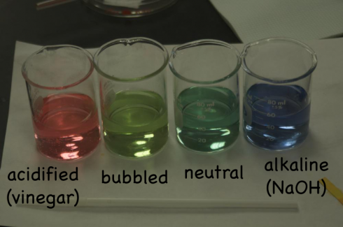
Changing colors of universal indicator show how blowing bubbles acidifies water (light green-second beaker) from neutral pH (dark green-third beaker) standard. For comparison, the first beaker (red) is acidified while the last beaker (blue) is made alkaline.

CO2 + H2O —-> H2CO3

This useful little reaction, where carbon dioxide reacts with water to produce carbonic acid, came up in my middle school class when we talked about respiration, it’ll come up soon in environmental science with the effects of carbon dioxide on the oceans (acidification), and it offers the opportunity to discuss pH and balancing chemical reactions in chemistry.

The middle school class did the neat little experiment where students blow bubbles in water (through a straw), and the carbon dioxide in their breath reacts with the water to slightly acidify it. A little universal pH indicator in the water (or even cabbage juice indicator) shows the acidification pretty well if you make sure to keep a standard nearby so students can see the change in color.

The fact that the CO2 in your breath is enough to acidify water begs the question — which was asked — how much of the air you exhale is carbon dioxide? According to the Oak Ridge Carbon Dioxide Information Analysis Center’s FAQ page, it’s concentration is about 3.7% by volume. Which is a lot more than the 0.04% average of the atmosphere.

Of course if you really want to talk about the pH you need to get into the acid equilibrium and the dissociation of the carbonic acid to produce H+ ions; you can get the these details here.

Flowers are “Creepy”

My students are researching the organisms they collected from the creek, and I was outlining the types of information I wanted them to find. We were talking about how many animals have seasonal reproductive cycles, and I pointed out that plants flower seasonally as well. One of my students put two and two together and came up with something close to a whole number: “You mean to say that flowers are … some sort of … creepy … sexual things?”

Microbe from the Creek

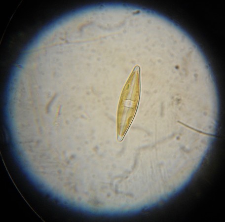
Microbe collected from the TFS Creek on 9/10/2012. Possibly a species of desmid.

The TFS campus has an excellent ecological gradient. It starts at the hydrologic base-level, with the small, usually permanent, creek in the valley. Then the landscape ranges up, past a narrow but dense riparian zone to the anthropomorphic campus, then up a shrub-covered hillslope that transitions abruptly into the advancing, mature, forest of the hill-top nature reserve. My environmental science class is taking advantage of our geographic proximity by doing a year-long ecological survey project.

We’ve just started, this fall, on the stream and riparian zone. I asked each of them to identify and do some research on a single organism. They all chose some type of macro-organism: spiders, crayfish, flowering herbs (note: just because it’s called an herb does not mean it’s edible), mushrooms, and more. There’s quite a bit of biodiversity down there, although, with the creek just now coming back from our particularly dry summer, the fish are few and far between.

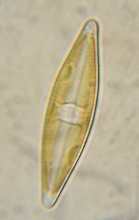
Close-up view of the micro-organism under 1000x magnification (oil immersion lens).

Since no-one chose to look for micro-organisms — even though I did suggest they were an important part of the ecology — I decided do so myself.

I found a loosely held together patch of algae, which I collected with the hope that it would harbor its own little microscopic ecological system. And it did. There were amoebas zipping around, the filamentous algae itself, and these little organisms that I can’t quite identify yet. T

hey may be desimids, but I’m not sure. They look slightly green, but I can’t see any clear chloroplasts (like these). I’ll try staining them tomorrow to see if I can identify any organelles.

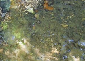
A terrible picture "showing" the patch of fillamentous algae I collected from the creek.