A nice animation showing the inner workings of a cell. There is a narrated version.
Tag: cells
Meiosis: Passing on Half of Your Genes
Now that we have an idea of what a strand of DNA looks like we’re going to start talking about how our genes are passed on to our kids.
During normal time (interphase) our DNA is stored in the nucleus of our cells. Humans have 23 pairs of chromosomes. Of each pair, one comes from your mom and one from your dad.

When a cell is not reproducing (which is most of the time) the chromosomes are unspooled threads in the cell’s nucleus.
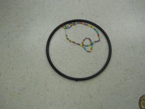
When the cell is preparing to reproduce, each DNA strand duplicates.
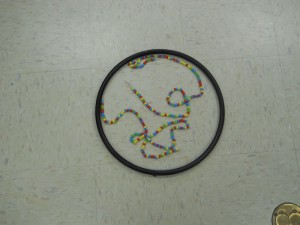
Then they fold up into the chromosomes and line up in the center of the cell.
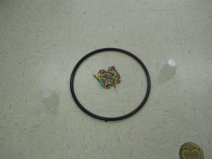
Now this is where interesting things start to happen. In mitosis, each chromosome pairs up with its duplicate, so when these are pulled apart you get two new cells with exactly the same DNA.

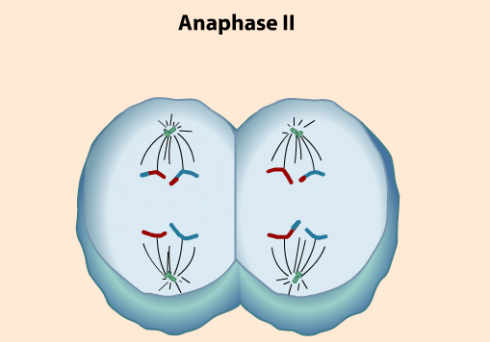
In meiosis however, where the cell breaks apart into reproductive cells called gametes, the two parent chromosomes pair up and exchange some DNA before being pulled apart (the DNA exchange is called crossing over). Since the DNA has duplicated before this happens, when the cell splits, you end up with two new daughters with mixed up DNA. Each daughter nucleus has two chromosomes, like all your other cells, but unlike every other (non-reproductive) cell in your body those chromosomes are different because of the DNA mixing. In addition, in the last step of meiosis (called Meiosis II) each daughter cell splits apart into two more daughter cells (granddaughter cells?) each with only one chromosome.

Again, it’s important to note that because of the crossing over and the second splitting, when everything is done, you end up with four cells — called gametes –, each of which has its own unique DNA. And unlike the other cells in your body, which have 23 pairs of chromosomes, each gamete only has 23 chromosomes.
Because a normal cell has 23 pairs of chromosomes is called a diploid cell. The gametes with only 23 single chromosomes is called haploid. These haploid gametes are the reproductive cells — eggs and sperm.
Thus, the DNA you contribute to your kids is not the same strands that you have in your cells, but a halved mixture of the two sets of genes you got from your parents.
References
The NIH has an excellent primer called “What is a Cell” on the history of cells, their parts, and how they split.
Meiosis
Going over meiosis in class today I used two videos. The first one was a bit simple. The second contained perhaps too much detail, but I showed it twice and stopped it at a few points on the second showing to point out the differences between mitosis and meiosis. I particularly wanted to highlight how genes are shuffled so the resulting reproductive cells have very different DNA from their parent. The shuffling is important because we’ll be comparing the advantages and disadvantages of asexual versus sexual reproduction later this week, as well as using Punnet squares to talk about heredity.
The first video:
The second:
Biology Videos
Sumanas, Inc. has an excellent collection of biology-related videos, including good coverage of mitosis and meiosis.
Unidentified Microbes (Gastrotrichs?)
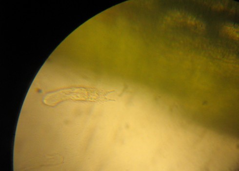
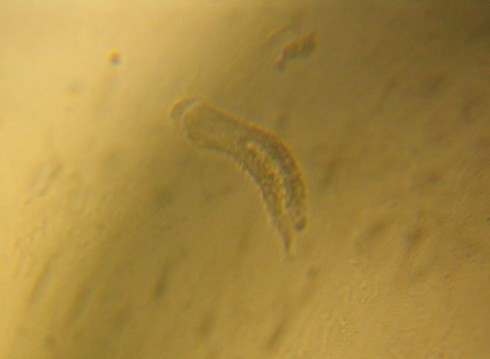
Update: I stumbled across this nice beginner’s guide to pond microbes that makes me thing these microbes are Gastrotrichs.
The Gastrotricha World portal has more information, as does the EOF and micrographia.
A video of a gastrotrich is down below.
—-
I’ve collected a set of aquatic plants for our fish tank for the middle school students to be able to look at their cells under the microscope. A few are from the store, like the Eregia densa I’ve used in the past, but we’ve also grabbed some algae from the creek, and Mr. Woodbury brought in some algae specifically for our two resident tadpoles.
I was checking out at the creek algae under the microscope when I came across these two microbes. They both were motile and seemed to be surrounded by cilia, but I really don’t know what they are.
Gene expression video
A short (abbreviated) introduction to gene expression — how proteins are produced from DNA.
From a Grain of Rice to a Carbon Atom

The Genetic Science Learning Center (which I’ve mentioned before) has a wonderful slider-bar animation that shows the differences in scale from what we perceive (a grain of rice or a coffee bean) down to the scale of cells, molecules and finally a carbon atom.
The smallest objects that the unaided human eye can see are about 0.1 mm long. That means that under the right conditions, you might be able to see an ameoba proteus, a human egg, and a paramecium without using magnification. …
Smaller cells are easily visible under a light microscope. It’s even possible to make out structures within the cell, such as the nucleus, mitochondria and chloroplasts. [my example] … The most powerful light microscopes can resolve bacteria but not viruses.
To see anything smaller than 500 nm, you will need an electron microscope. … The most powerful electron microscopes can resolve molecules and even individual atoms.
–Genetic Science Learning Center (2011, January 24) Cell Size and Scale. Learn.Genetics. Retrieved June 13, 2011, from http://learn.genetics.utah.edu/content/begin/cells/scale/
Viruses in Glass
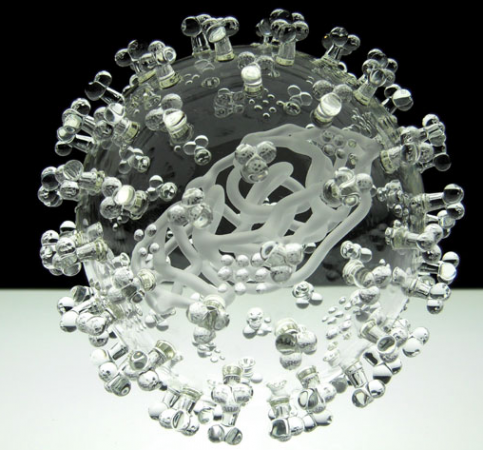
Some people knit bacteria, our students create models of plant and animal cells as an assignment, and Luke Jerram blows viruses in glass.
Interestingly, he does not use colors, just clear, transparent glass, because while almost all pictures you see of viruses, from scanning electron microscopic images to text-book diagrams, have color, the color is added for scientific visualization. So, he believes, his glass replicas are more realistic. This is described in this interview.
About the works:
The pieces, each about 1,000,000 times the size of the actual pathogen, were designed with help from virologists from the University of Bristol using a combination of scientific photographs and models.
— Caridad (2011): Harmful Viruses Made of Beautiful Glass
The video below shows how it’s done.
There is a lot more information on the art and the science at the Glass Microbiology website.