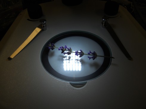
Lavender Flowers Up Close

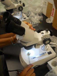
Lavender flowers on the stage of the reflecting, stereo microscope.

In addition to the basic stereoscopes with their fixed 10x and 30x magnifications, we also acquired a zoom stereoscope for more serious research projects. I tried it out with a sprig of lavender blossoms.
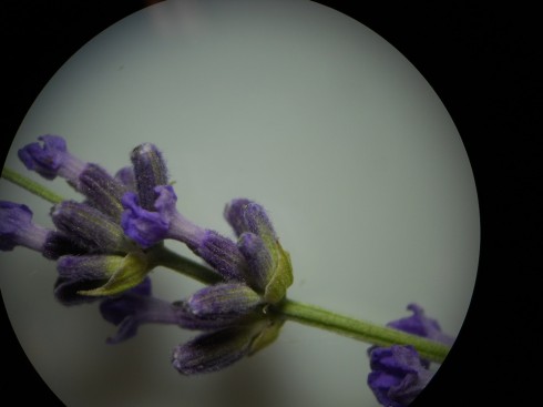
Closeup of lavender flowers. Magnification 7x.

The clips on the stage weren’t particularly useful for holding something as small as a single flower, so, to see into the flower, I had to improvise with some of the dissection gear.
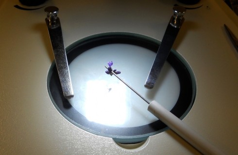
Holding the lavender flower upright on the stage with a dissection probe.

At larger magnifications, the focal depth is pretty small so it’s tricky trying to get the big picture. Even thought the camera didn’t quite capture it, you can make out the pollen grains.
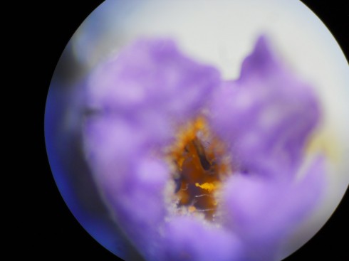
Looking into a lavender flower from the top. Magnification ~45x.

I tried slicing the flower longitudinally to get a better look inside, and to see how difficult it would be to identify the major parts.
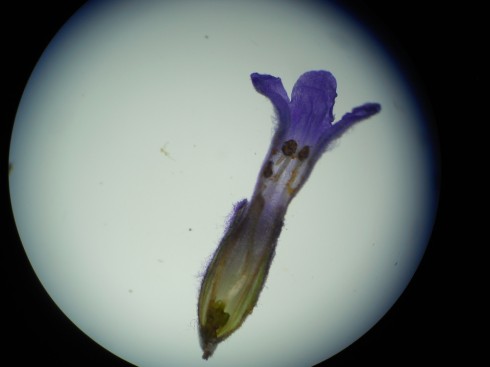
Longitudinal section of a lavender flower. Magnification 14x.

The photos turned out well using a point-and-shoot Nikon camera through the eyepiece, but even these pictures did not capture all the detail visible to the eye.
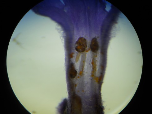
Lavender flower sliced longitudinally. Two stamens are clear visible. Magnifications ~50x.

With the 2x objective attached, the microscope gets up to 90x magnification, but it becomes very hard clearly see anything after about 60x. All in all, the optics are good, and the lights bright enough to make for a very nice microscope.
Green Onion Under the Microscope

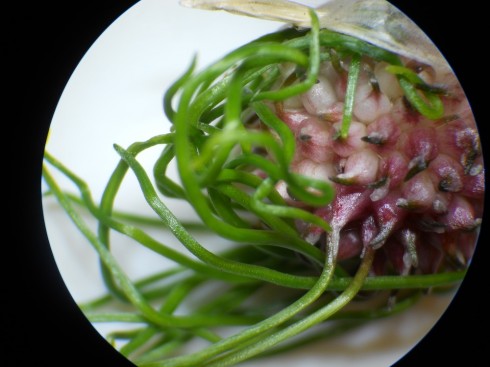
Seed head of a green onion. 10x magnification.

A new set of stereo, reflected-light, microscopes came in last week, and I’ve been testing them out. MPU has a good eye for these things, so I asked him to collect some samples for examination.

The first thing he came up with was this beautiful green onion. The seed head has some remarkable colors, and the microscopes are of good enough quality that we could examine in quite good detail at 10x magnification. We were even able to see a few small insects hanging out on the seed head that would have been invisible to the naked eye. They didn’t like the light, however, and hid before I could get a good photo.

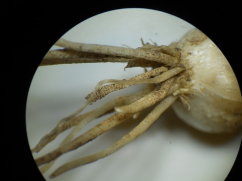
Roots of a green onion. 10x magnification.

Life Under the Ice

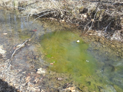
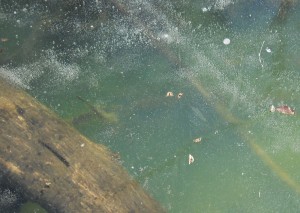
Algae growing under the ice on the creek.

While not quite as dramatic as drilling through four kilometers of ice to find signs of live in an Antarctic lake that’s been isolated from the rest of the world for over 100,000 years, we observed filamentous algae blooming under the clear ice on the creek earlier this winter.

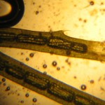
Algae under the microscope (40x magnification).

I collected some of the algae and put it into the fish tank; I was hoping I could use it when we looked at plant cells because aquatic plants tend to have larger cells that are easier to see under the microscope.

However, one of my students is keeping tadpoles (also from the creek) in the fish tank. She noticed that the tadpoles were hanging out on top of the algae, and the algae was disappearing. Well, at least we’d solved her problem about what to feed the tadpoles.

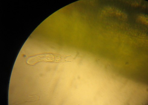
Unknown microbe hanging out in the algae.

While the filamentous algae might not be as good as the Egeria densa for plant-cell microscopy, it does host quite a number of other microbes that are fascinating to look at.

Ecological Role of Algae

Based on these observations, ecologically the filamentous algae does not just provide habitat for protists and other microbes, it also appears to be a significant source of food for larger animals, like the tadpoles, and probably also the small fish that live in the creek.

Bubbles trapped under the ice. With all the algae growing in the water, and the clear ice, these bubbles may well be made of oxygen.

Therefore, I’d hypothesize that in the winter, when the fish disappear, and most of animal life is quite subdued, the algae blooms because it’s not being grazed on nearly as much (see the picture above). When the weather warms, however, it’s the turn of the algae to repressed.

It would be interesting to have a student monitor the algae growth, and the fish/tadpole population, over the course of the school year to see if the relationship is more than just coincidence.

P.S. After our last snowfall, the melting snow has put a lot of water into the creek, and all the algae appears to have been washed away.

Unidentified Microbes (Gastrotrichs?)

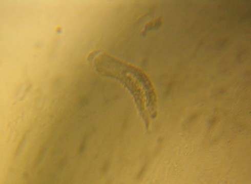
Unknown microbe hanging out in the algae. (100x magnification)

Update: I stumbled across this nice beginner’s guide to pond microbes that makes me thing these microbes are Gastrotrichs.

The Gastrotricha World portal has more information, as does the EOF and micrographia.

A video of a gastrotrich is down below.

—-

I’ve collected a set of aquatic plants for our fish tank for the middle school students to be able to look at their cells under the microscope. A few are from the store, like the Eregia densa I’ve used in the past, but we’ve also grabbed some algae from the creek, and Mr. Woodbury brought in some algae specifically for our two resident tadpoles.

I was checking out at the creek algae under the microscope when I came across these two microbes. They both were motile and seemed to be surrounded by cilia, but I really don’t know what they are.

Unknown microbe number 2 (100x magnification).

Microbe from the Creek

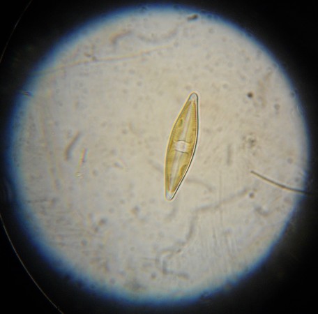
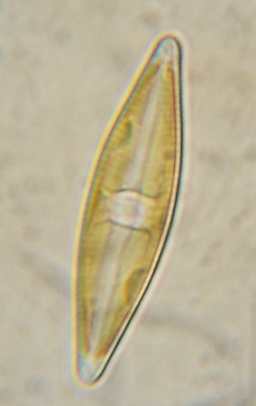
Microbe collected from the TFS Creek on 9/10/2012. Possibly a species of desmid.

The TFS campus has an excellent ecological gradient. It starts at the hydrologic base-level, with the small, usually permanent, creek in the valley. Then the landscape ranges up, past a narrow but dense riparian zone to the anthropomorphic campus, then up a shrub-covered hillslope that transitions abruptly into the advancing, mature, forest of the hill-top nature reserve. My environmental science class is taking advantage of our geographic proximity by doing a year-long ecological survey project.

We’ve just started, this fall, on the stream and riparian zone. I asked each of them to identify and do some research on a single organism. They all chose some type of macro-organism: spiders, crayfish, flowering herbs (note: just because it’s called an herb does not mean it’s edible), mushrooms, and more. There’s quite a bit of biodiversity down there, although, with the creek just now coming back from our particularly dry summer, the fish are few and far between.

Close-up view of the micro-organism under 1000x magnification (oil immersion lens).

Since no-one chose to look for micro-organisms — even though I did suggest they were an important part of the ecology — I decided do so myself.

I found a loosely held together patch of algae, which I collected with the hope that it would harbor its own little microscopic ecological system. And it did. There were amoebas zipping around, the filamentous algae itself, and these little organisms that I can’t quite identify yet. T

hey may be desimids, but I’m not sure. They look slightly green, but I can’t see any clear chloroplasts (like these). I’ll try staining them tomorrow to see if I can identify any organelles.

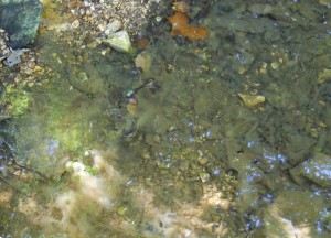
A terrible picture "showing" the patch of fillamentous algae I collected from the creek.

Spittlebugs

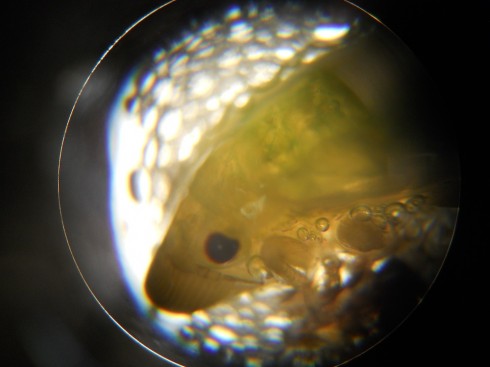
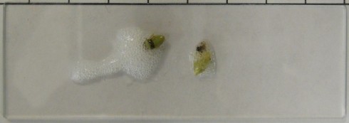
40x magnification of the head of the spittlebug nymph.

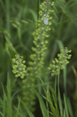
On the wildgrass-covered slope next to school, you can see a lot of these little foamy things, that look like spit, on the stalks of the tall grasses and herbs.

Spittlebug "spit" is mostly made of a froth of the plant's sap.

One of my students collected some to look at under the microscope. We thought it might be the collection of eggs of some creature. It turned out that, at the center of the foam, was what looked like an immature insect. A quick google search for “spit bugs” turned up froghoppers, whose nymphs create the spit to protect them from the environment (heat, cold) and hide themselves from predators.

They suck the sap of the plants they’re on, and can be agricultural pests.

Spittlebug nymphs on a slide.

Guide to Using a Microscope

Sitting innocuously on the clearance table at a Barnes & Noble (in Cedar Rapid, Iowa actually) was a copy of Georg Stehli’s The Microscope and How to Use It.

At 75% off it was less than $3, which is quite a steal for a guide to what I found to be the most fascinating piece of scientific equipment for my middle schoolers. One of their first natural world lessons was on how to use the microscope. In the classroom there was always one sitting on the shelf, protected by its translucent plastic cover, but easily accessible.

I also took one everywhere, including to the cabins on our immersion trips, which is where they discovered the crystalline structure of salt and sugar grains, and the microfossils at Coon Creek.

And, interestingly enough, my microscopy posts are some of the most popular posts on this blog (the onion cell is regularly in the top ten).

The Microscope and how to use it by Georg Stehli.

Apart from the basics of how to use a microscope, Stehli’s book goes into simple sample preparations and preservation for almost everything you’re likely to encounter in the curriculum, in the classroom, and in the back yard. Though neither crystal structure nor microfossils are covered, the techniques for looking a the hard parts of biological specimens are applicable.

I would have loved to have had a copy of this last year when I was trying to figure out which were the best dyes to use for some of the odder samples my students came up with, and how to make them into permanent slides. It’s not easy to find this kind of broad reference online.

Salt and Sugar Under the Microscope

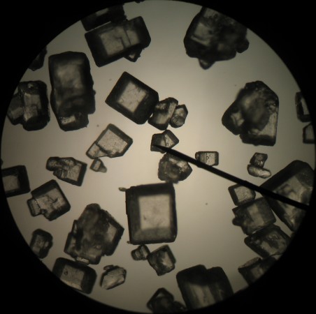
Sugar crystals under 40x magnification.

Salt and sugar crystals have wonderfully distinctive crystal forms. They might well be good subjects for introducing minerals, crystals and some of the more complex geometric solids.

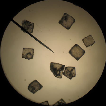
Cube shaped salt crystals under 40x magnification.

The salt crystals are clearly cubic, even though some of the grains seem to be made up of overlapping cubes.

The atoms that make up salt's atomic lattice are arranged in a cubic shape, which results in the shape of the salt crystals. The smaller grey atoms are sodium (Na), and the larger green ones are chlorine (Cl).

Salt is an ionic compound, made of sodium and chloride atoms (NaCl). When a number of these molecules get together to form a crystal, they tend to arrange themselves in a cubic pattern. As a result, the salt crystals are also cubic. In fact, if you break a salt crystal, it will tend to break along the planes that are at the surfaces of the planes of the atomic lattice to create a nice, shiny crystal faces. Gem cutters use this fact to great effect when they shape diamonds and other precious stones.

Of course different crystals have different atomic arrangements. The difference is clear when you compare salt to sugar.

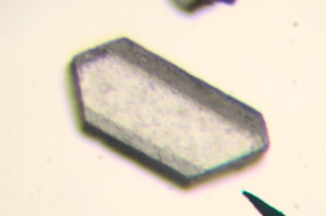
A single sugar crystal looks a bit like a fallen column.

Sugar crystals look a bit like hexagonal pillars that have fallen over. According to the Beet-sugar handbook (Asadi, 2007), sugar crystals actually have a monoclinic form, which could end up as asymmetric hexagonal pillars. Salt crystals, on the other hand, have the habit of forming cubes.