Crayfish: Charismatic Mesofauna

This year, the creek is teeming with crayfish, especially compared to last year during the drought when the creek dried up and the crustaceans were hard to find. I had five students out collecting organisms on Wednesday, and they came back with ten crayfish ranging in size from a couple centimeters long, to one that was about 12 centimeters from claws to tail.

Crayfish in a jar.
Crayfish in a jar. Seen through stereoscope. Magnification unknown.

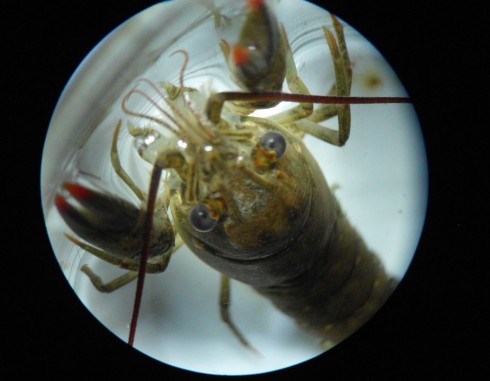
I was just looking at one of the pictures I took and realized that I did not know what species it belonged to. I’ll be having students do reports on individual species for biology next year, and I’d be very surprised if someone did not choose crayfish. They’re so many of them and, as my students from Wednesday will attest, they’re just so charismatic. While I’ve not looked into it much myself, the Crayfish & Lobster Taxonomy Browser seems a decent place to start researching.

Stomata of a Basil Leaf

It’s not always easy to see stomata, the pores on the surfaces of leaves that allow plants to breathe. I tried the leaves of pepper and tomato plants with a dissecting microscope to no avail. However, compared to these, the stomata on basil leaves were enormous. They were actually visible on the lowest magnification (6x), but the higher magnification is necessary to make out the detail.

For these pictures, I was lucky enough to have gotten to try out one of Leica’s research grade, digital microscopes (the DMS1000b). Given that I only had a hour or so, I did not take the time to experiment with all the optimization options, however, the pictures turned out remarkably well, none-the-less. In particular, you might note the highest magnification images, labeled 48(d)x, are just digital enlargements from the 48x magnification pictures.

Having the built in camera makes it an awful lot easier to put images into the online microscope app, and I suspect will make focus stacking a lot easier as well. Since this scope is a bit out of the range of my small high-school budget, I really need to figure out a good, solid way of mounting my point-and-shoot cameras on the microscopes I have.

Ticks in the Woods

While walking through the woods to recover the skeleton the other day, I picked up, or rather was boarded by, a few ticks. So when I got back to school I plopped them under the stereoscopes to try to identify them.

Lone star tick (Amblyomma americanum) (adult female?) from the woods behind Maggie’s house (Missouri). Magnification ~20x; dorsal view.

They were both lone star ticks (Amblyomma americanum): one adult and one juvenile.

Lone star tick nymph (magnification 45x; dorsal view).

Under the microscope, they were quite pretty with their very interesting red and black patterns.

Note:

Purdue’s extension service has an excellent reference page on basic tick anatomy and ecology for a lay audience, and Colonial Pest Control has a very readable overview of The Biology of Ticks ( Jay O.) that goes into their life cycles and how to deal with them.

Much greater detail can be found in the Tick Gross Anatomy Ontology, but the best reference I’ve found so far is the USDA’s Handbook (485): Ticks of Veterinary Importance (pdf). There you can find great anatomy diagrams and interesting biological and ecological information. One curious piece of information is that ticks can survive a long time (three years in one case) without a blood meal. It also includes some excellent diagrams:

Diagrams of lone star tick external anatomy from USDA Handbook 485: Ticks of Veterinary Importance.
External anatomy of hard bodied ticks. From the USDA Handbook 485: Ticks of Veterinary Importance.

A Catalpa Tree Flower Under the Microscope

More testing of the higher powered stereo-microscope with this flower specimen from a catalpa tree on campus.

Anthers with pollen grains (~25x).

The catalpa tree leaf and flower for reference.

Large catalpa leaf and two flowers.
Detail of longitudinally bisected flower (~7x).
Closeup of petal (~35x).
The colors on the petals come from cells having different colors (~90x).

Lavender Flowers Up Close

Lavender flowers on the stage of the reflecting, stereo microscope.

In addition to the basic stereoscopes with their fixed 10x and 30x magnifications, we also acquired a zoom stereoscope for more serious research projects. I tried it out with a sprig of lavender blossoms.

Closeup of lavender flowers. Magnification 7x.

The clips on the stage weren’t particularly useful for holding something as small as a single flower, so, to see into the flower, I had to improvise with some of the dissection gear.

Holding the lavender flower upright on the stage with a dissection probe.

At larger magnifications, the focal depth is pretty small so it’s tricky trying to get the big picture. Even thought the camera didn’t quite capture it, you can make out the pollen grains.

Looking into a lavender flower from the top. Magnification ~45x.

I tried slicing the flower longitudinally to get a better look inside, and to see how difficult it would be to identify the major parts.

Longitudinal section of a lavender flower. Magnification 14x.

The photos turned out well using a point-and-shoot Nikon camera through the eyepiece, but even these pictures did not capture all the detail visible to the eye.

Lavender flower sliced longitudinally. Two stamens are clear visible. Magnifications ~50x.

With the 2x objective attached, the microscope gets up to 90x magnification, but it becomes very hard clearly see anything after about 60x. All in all, the optics are good, and the lights bright enough to make for a very nice microscope.

Green Onion Under the Microscope

Seed head of a green onion. 10x magnification.

A new set of stereo, reflected-light, microscopes came in last week, and I’ve been testing them out. MPU has a good eye for these things, so I asked him to collect some samples for examination.

The first thing he came up with was this beautiful green onion. The seed head has some remarkable colors, and the microscopes are of good enough quality that we could examine in quite good detail at 10x magnification. We were even able to see a few small insects hanging out on the seed head that would have been invisible to the naked eye. They didn’t like the light, however, and hid before I could get a good photo.

Roots of a green onion. 10x magnification.