How Microscopic Shells can tell us the History of the Earth’s Climate

Seeing the bigger picture.

Looking at the smear slides of Coon Creek Sediment Matrix got me thinking about just how important these little, microscopic shells have been for what we know about the Earth’s past climate. In fact, they provide the background knowledge that we have about the changes in climate that we’re seeing today.
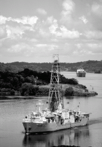
Deep sea drilling vessel, JOIDES Resolution. Image via the National Science Foundation.

Back in the 1970’s the Deep Sea Drilling Project collected a lot of sediment cores from all around the world. The deeper you drill under the sea bed the older the sediments are, so micropaleontologists could look at how the organisms that lived in a certain area changed over time. Certain forams that could only live in warm oceans were found living far to the north. By combining all the information from all the sediment cores, they could construct paleo-geographic maps showing what the climate was like in the far past. It’s one of the reasons we know that the Jurassic climate was a lot warmer than today’s climate.

Then they invented mass spectrometers.

Mass specs can find the mass of individual atoms. Calcium carbonate has the chemical formula CaCO3. Water, as we should know by now, is H2O. They both have oxygen atoms, but not all oxygen atoms are equal; some are more equal. Actually, the mass of any atom is made up of the mass of the protons plus the mass of the neutrons in its nucleus. Now, by definition, any atom with eight protons is oxygen; however, while oxygen usually has eight neutrons, it sometimes has nine or even ten.

Your standard oxygen, with eight protons and eight neutrons has an atomic mass of sixteen, and is written as 16O or oxygen-16. Well, oxygen with ten neutrons is going to have a mass of eighteen (8p + 10n) and be called oxygen-18 (18O). These different versions of the same element are called isotopes.
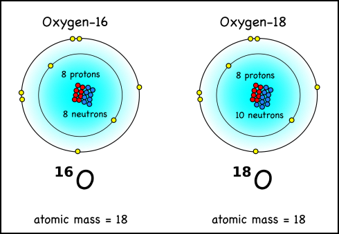
Oxygen-18 has two more neutrons than the much more common oxygen-16. Note that both atoms have eight electrons, but their masses don't count because electrons are really small compared to the protons and neutrons which have about the same mass.
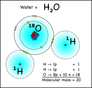
Water molecule with a molecular mass of 20.

What does this have to do with climate? Well a water molecule with two hydrogen atoms, each weighing one atomic mass unit, and one oxygen-16 atom will have a molecular mass of 18, while a water molecule with an oxygen-18 atom will have a mass of 20. When water evaporates from the oceans, the water with the lighter isotope will have an easier time going from liquid to a gas in the atmosphere.

So, during an ice-age for instance, lots of water evaporates from the oceans, falls on land as snow, and then gets trapped in the enormous glaciers that cover entire continents. Since the lighter water molecules evaporate easier from the oceans, they’re the ones that will end up falling as snow and being compressed into glacial ice. The water molecules left behind in the ocean will tend to have the heavier oxygen-18 isotopes. Since the forams use the ocean water as part of the process of creating their calcium carbonate shells, the oxygen from the water ends up in the carbonate (CO3) of the shells. Since the ocean water has extra oxygen-18s during an ice-age, then the shells will have extra oxygen-18 isotopes during an ice-age.
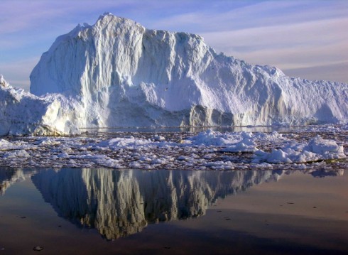
Ridge of ice from the continental glacier in Greenland. Glacial ice will have lighter isotopes than the oceans the water originally evaporated from.Image by Konrad Steffen from the U.S. Antarctic Survey.

Therefore, by measuring the amount of heavy oxygen-18 isotopes that are in a single shell, we can tell how large the glaciers were at the time that shell formed, and tell what the global climate was like.

Of course there are some interesting complexities to the story, but that’s the general idea of how the microscopic shells of long-dead plankton can tell us about the history of the Earth’s climate.
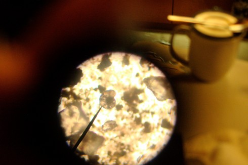
Coon Creek Matrix Under the Microscope

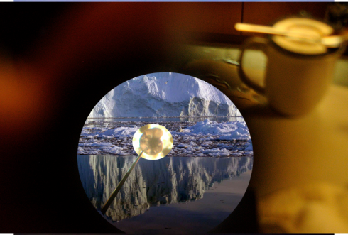
Hunting for microscopic fossils at the dinner table. Inside the circle is 100x magnification; outside the circle the magnification is 1.

My students will tell you that I’m never happier than when I have my cup of tea. On the night after our visit to Coon Creek, I put a tiny sample, about the size of a matchstick’s head, of sediment matrix on a microscope slide, and added a drop of water to disperse the grains. Then I sat there, while the chaos of dinner-making swirled around me, and searched for tiny, microscopic fossils of creatures that died long ago. With my cup of hot tea beside me, it was like sitting in the eye of a storm, flaming hamburgers be damned, a modicum of sanity in the asylum.

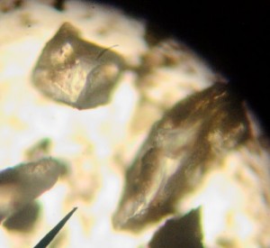
Quartz grains from Coon Creek Formation sediment seen under the microscope at 100x magnification. Quartz is easy to identify because of the way it breaks with curved fractures.

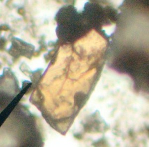
The first thing I noticed though were the quartz grains. They’re very small, silt-sized, but are the largest grains in the sediment. They’re pretty easy to identify because they break like glass, with curved, conchoidal fractures. They’re also pretty little things under the microscope; little, sharp-peaked, transparent mountains.

Other minerals are visible in the sediments. Though they're relatively large they're still dwarfed by the quartz (100x magnification).

Other minerals are visible in the slides, but they’re dwarfed in size and quantity by the quartz. Yet there is enough of the dark green, glauconite clay to bind the quartz grains together and protect the shells embedded in the sediment from dissolution by the universal solvent, water.

It’s interesting to observe these other minerals, because they take the more classic crystalline shapes and forms. The sharp edges are parallel to one another because of the alignment of the atoms in the mineral crystal.

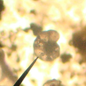
Snail like shape of what's probably a planktonic (lives in the water) foram. (100x magnification).

Finding the micro-fossils took a little patience. The entire slide had only four obvious specimens. Since they’re so small that meant a lot of going back and forth under the small field of view of the 100 magnification objective lens. They look like foraminfera to me, but it’s been a while since I’ve encountered them. Foraminfera, or forams for short, are tiny organisms that secrete beautiful calcium carbonate shells. They can be found in, or in the sediments beneath, most of the world’s oceans, particularly in the warmer areas.

Finding forams in the Coon Creek Matrix is a nice little exercise. One of my students, seeing what I was doing, wanted to try it too, so she made her own slide and searched until she found her own specimen. It was somewhat inspiring, so I’ve put together a more detailed post about finding microfossils.

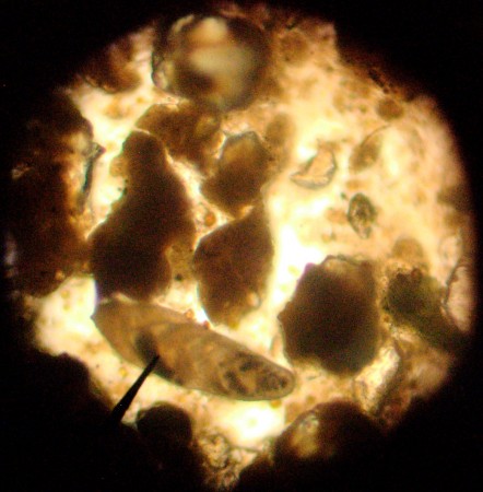
We also found a neat little shell that looks like the overlapping scales on a pine cone. We were disconnected from the internet, so I was only able to look it up when I got back to school.

What looks like a type of boliviana foraminfera. It's benthic, which means that it lives in the sediments not in the water.
What looks like a type of bolivina foraminfera. It's benthic, which means that it lives in the sediments not in the water. (100x magnification).

Dr. J Bret Bennington at Hofstra has posted a nice PowerPoint of his introduction to marine microfossils lecture. As a basic introduction, it’s quite comprehensible to middle-school students, or people like myself who did not pay as much attention as they should have during that part of Paleontology. Anyway, based on these notes, the pine-cone-shaped thing is probably a variety of bolivina, a benthic foraminfera. The Foraminifera.eu-Project, is a wonderful, volunteer-produced resource for pictures and identifying forams.

Bolivina are benthic, which means they spend most, if not all of their time in the mud. Planktonic micro-organisms, on the other hand, spend their lives floating around in the water.

Foraminfera have calcium carbonate shells, as do clams and oysters. In the shallow oceans there is a slow rain of them that cover the sea-bed over the millenia. You can end up with thick layers. In fact, the white cliffs of Dover are white because of all the microscopic calcium carbonate shells. In the deeper reaches of the oceans there are much fewer of these shells because they dissolve under high pressure. As a result, down there you tend to find microfossils of diatoms and radiolarians, things with silica shells. Silica is that same material from which glass is made, and is the same material in quartz.

Finding microfossils has actually been quite important for understanding the history of the Earth’s oceans and climate. But that’s another story.

Cells, cells, cells

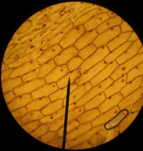
Onion cells stained with iodine. 100x magnification.

We spent the afternoon period on science. I’d given some individual microscopy lessons during the last immersion, where we looked at exciting protozoans moving around in pond water. This time they tried their hands at onion cells and staining with iodine, using a very nice and clear YouTube video posted below (kyliefansunited, 2008) as a reference.

Nucleus of an onion cell stained with iodine and, for experimentation, Congo Red. 1000x magnification

The immersion oil had arrived in the mail earlier in the week so we got to try out the 100x oil lenses. We can now see structures inside the nucleus quite nicely.

Other things did not go so well. I’d written up, using the excellent recommendation of Anna Clarke, what I though was a neat exercise to look at the effect of osmosis on the cells of a waterplant, Egeria densa. The small group struggled with it, I think in large part because they were not quite prepared (had not done the background reading), and weren’t working very well together today. I’ll keep it on the schedule, but next time I’ll have to think hard on if it will be necessary to tweak the exercise.

Osmosis under the microscope

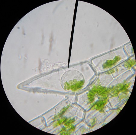
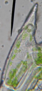
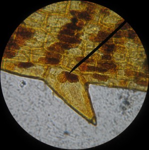
The effects of placing freshwater plant cells (Egeria densa) in salt water solution.

In a bit of a hurry, I swung by the pet store and picked up the aquatic water plant with the thinnest leaves I could find. It turned out to be Egeria densa, and while not the Elodea recommended by my expert contact Anna Clarke as a good subject for some microscope work, it seemed quite similar.

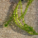
Egeria densa plants sitting in shallow water in the sun.

I needed the plant for an osmosis experiment. Dropping a little salt water on leaf cells of a freshwater plant should suck all the water out of the vacuoles and through the cell walls, potentially collapsing the cells (wouldn’t that be cool). I’d never done this before so I was quite curious to see what would actually happen.

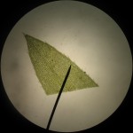
Leaf tip of Egeria densa. 40x magnification.

The leaves have multiple layers of cells, so it’s hard to distinguish much at the center of a freshly clipped leaf, especially at high magnification. But if you look at the cells at the edges of the leaves, you can see some really neat looking, spiky cells, for which, I’m willing to bet, biologists have some really cool, multisyllabic name.

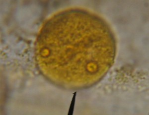
Spiky cell under 1000x magnification.

With a little bit of immersion oil and a 1000x objective, the spiky cells are good subjects for magnification: they’re a bit larger than their neighbors so they’re easier to see; their chloroplasts are distinct; and you can even make out the nucleus without staining.

Then I added the salt solution, and while the cell walls stayed strong, the cytoplasm collapsed into a little droplet at the center of the cell. The chloroplasts and the nucleus were all bundled together in this central blob (see the image at the top of the post). It’s quite the neat effect, though not exactly what I thought to see.

Spiky Egeria cells with iodine stain.

An interesting side note is that the cell nuclei show up very nicely with iodine stain, but the stain also discolors the chloroplasts.